|
|
Biography |
|
|
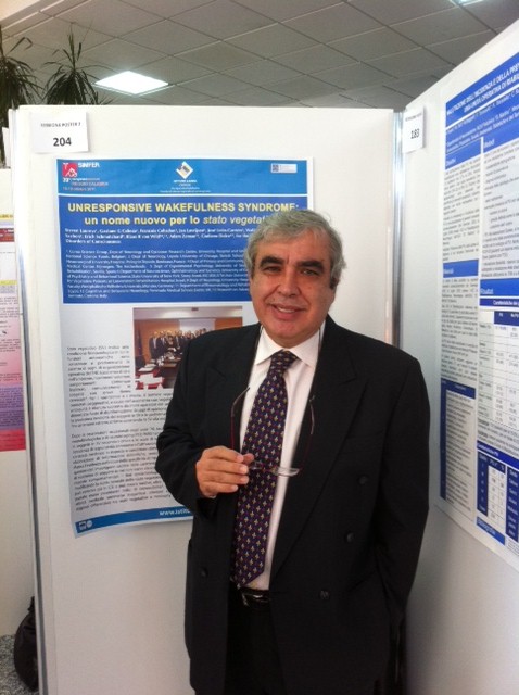
 Prof. José León-Carrión, Ph.D.
Education/career: BA in Philosophy, Ph.D. in Psychology, Universidad Autónoma de Madrid, Spain. Professor of Neuropsychology & Director of Human Neuropsychology Laboratory at University of Seville, Spain. Director of R+D+I Department at the Center for Brain Injury Rehabilitation (C.RE.CER.), Seville. Membership/Distinction: Executive Committee Vice Chair,International Brain Injury Association (IBIA; founding member, World Academy for Multidisciplinary Neurotraumatology; member, European Brain Injury Society and various journal editorial boards; reviewer and consultant,U.S. Department of Defense TBI Grant Program;recognized internationally for over two decades of work in rehabilitation and brain injury. Expertise:International expert, consciousness studies and rehabilitation of patients in coma, vegetative state, minimal conscious state, locked-in syndrome and severe neurocognitive disorders; investigator,studies on TBI rehabilitation; developed neuropsychological assessment tools: Computerized Sevilla Neuropsychological Test Battery, Luria’s Memory Words-Revised Test and Neurologically-related Changes of Personality Inventory (NECHAPI); developed rehabilitation for individuals with disabilities; author, textbooks and articles in neuroscientific journals and books. Recent books: “Neuropsychology and the Hispanic Patient”, “Behavioral Neurology in the Elderly”, “Brain Injury Treatments: Theory and Practices”, and “Evaluación Clínico Neuropsicológica de la Afasia Puebla-Sevilla”. Author and co-author,recent articles:
The Infrascanner, a handheld device for screening in situ for the presence of brain haematomas. Brain Injury, 2010; Unresponsive wakefulness syndrome: a new name for the vegetative state or apallic syndrome. BMC Med. 2010; Accuracy of the S100ß Protein as a marker of brain damage in Traumatic Brain
Injury. Brain Injury,In Press;
Time and course of functional Rehabilitation after deep traumatic coma. JRMed, In Press.
|
|
|
|
|
|
|
Abstract |
|
|
|
|
|
|
|
|
Humans and animals share a brain that differs little in terms of anatomy and function. However, these differences are what make us human, defining our species as unique in our universe. A quality unique to humans and primates is a granular frontal cortex in the frontal lobe. The prefrontal cortex grants us the ability to be independent and decision-making capabilities, while the posterior cortex contains any and all information which has entered our brain. In most mammals, prefrontal and parietal cortical areas have been identified as subserving sensory-motor, cognitive, emotional, motivational, behavioural, executive, and visceral functions. Yet, what sets humans apart is consciousness, a characteristic regulated by activation of the frontal-parietal network. The disconnection of this cerebral network leads to disorders of consciousness.
Two types of interacting cortical neurons are purposed to be involved in consciousness (Crick and Koch, 2003). One group is said to project from the posterior cortex, and the other from the anterior cortex. The anterior, or executive, cortex appears to be “looking at” the posterior, or sensory, cortical system. These two groups of neurons continuously interact via long and short cortico-cortical and cortico-thalamo-cortical routes to create consciousness (Crick and Koch, 2003). The temporal organization of stimuli in the brain requires serial and alternating engagement of frontal and posterior cortices (Leon-Carrion et al., 2006). Posterior cerebral regions allow humans and animals to function automatically, while prefrontal regions provide a purpose to human activity.
A large body of neuropsychological evidence supports the existence of these cortical areas, reinforcing the relevance of frontal lobe and retrolandic areas in the generation of consciousness. Regulation of the content of consciousness has been associated with the flexibility of prefrontal cortical functions, while various aspects of sensory processing have been linked to posterior cortical functions (Lhermitte, 1983; Passingham, 1993; Fuster, 2008). According to John (2005), awareness is reduced when the prefrontal cortex (PFC) is depressed. The specific sequences of neural links engaged in consciousness and awareness may be disturbed in patients with frontal lobe lesions [Luria, 1966; John, 2005; Leon-Carrion et al., 2008).
Survivors of traumatic brain injury (TBI) often suffer disorders of consciousness as a result of a breakdown in cortical connectivity. However, little is known about the neural discharges and cortical areas working in synchrony to generate consciousness in these patients. In this presentation, we analyzed cortical connectivity in patients with severe neurocognitive disorder (SND) and in the minimally conscious state (MCS). We found two synchronized networks subserving consciousness; one retrolandic (cognitive network) and the other frontal (executive control network). The synchrony between these networks is severely disrupted in patients in the MCS as compared to those with better levels of consciousness and a preserved state of alertness (SND). The executive control network could facilitate the synchronization and coherence of large populations of distant cortical neurons using high frequency oscillations on a precise temporal scale. Consciousness is altered or disappears after losing synchrony and coherence.
We suggest that the synchrony between anterior and retrolandic regions is essential to awareness, and that a functioning frontal lobe is a surrogate marker for preserved consciousness, a human navigation marker in the organization of complex goal-directed behaviour.
|
|
|
|
|