| front |1 |2 |3 |4 |5 |6 |7 |8 |9 |10 |11 |12 |13 |14 |15 |16 |17 |18 |19 |20 |21 |22 |23 |review |
 |
Anatomical
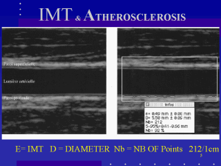
studies (1) showed that the distance between the two parallel
lines represents the IMT. The best site for measurement is the far wall of the distal common carotid artery, with a better reproductibility than at the bifurcation or on the internal carotid. It is also possible to measure IMT of the popliteal artery or of the common femoral. |