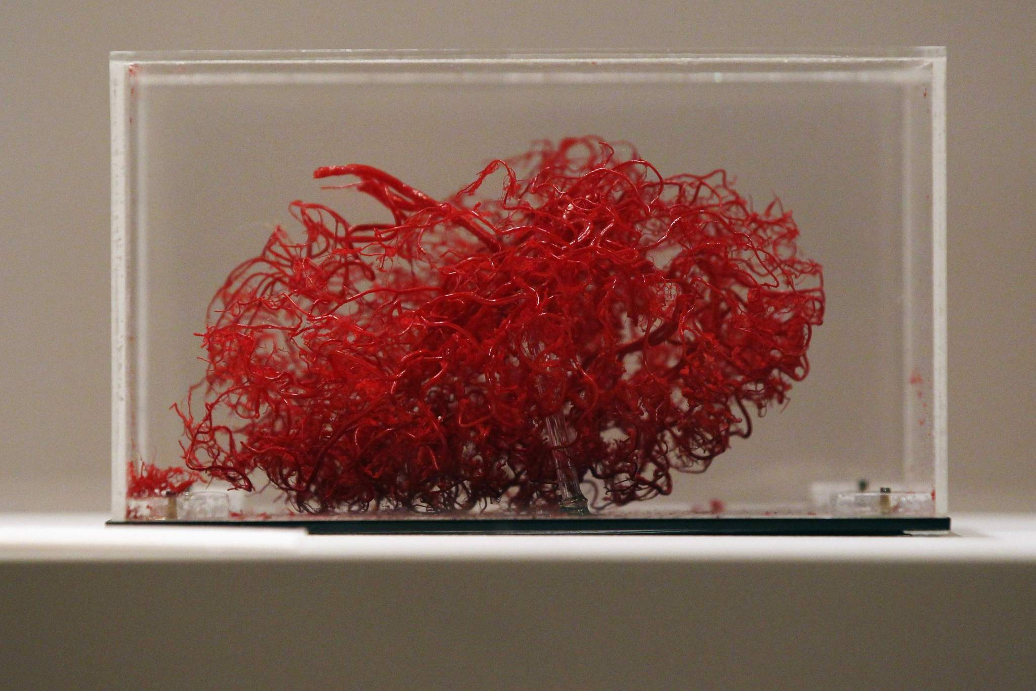
3D-Printed Vasculature Networks Created
Share


Technology is deeply entrenched in every aspect of our lives. Researchers in the field of 3D printing technology have been actively attempting to come up with new methods to artificially create lifelike blood vessels that can fully function in the human body.
Up until recently, 3D printing technologies have been slow, costly, and are only capable of producing single blood vessels; a tube that still does not integrate with the body’s own vascular system. However, that has changed; thanks to technology.
Nanoengineers at the University of California, San Diago, led by Dr. Shaochen Chen, a nanoengineering Professor, have dived into a major challenge in tissue engineering. They have managed to 3D-print lifelike tissues and organs with functional vasculature, which are networks of blood vessels that can transport blood, nutrients, waste, and other biological materials, and perform those functions safely when integrated in the human body.
Human tissues and organs require blood vessels to survive and function properly, and this is where organ transplants, which are high in demand and short in supply, become problematic. The technology boost in the science of tissue engineering can be the reason for great change. Chen’s lab has artificially created a vasculature network that can safely implant with the body’s own vessel network to circulate blood. These blood vessels then branch out into much smaller vessels, similar to the blood vessel structure in the body.
The team of scientists developed an innovative bioprinting technology, using their own homemade 3D printers, to rapidly produce 3D microstructures that act like the sophisticated designs and functions of biological tissues. Chen’s lab has made use of this technology in the past to create live tissue and microscopic fish able to swim in the body and remove toxins.
A lot of effort goes into the creation of this tissue. First, a 3D model of the biological structure is designed on a computer. The computer then transfers 2D snapshots to millions of microscopic-sized mirrors, which are each digitally controlled to project patterns of UV light in the form of these snapshots. The UV pattern is then exposed to a solution containing live cells and light-sensitive polymers that solidify upon exposure to UV light. The structure is then printed layer by layer, creating a 3D solid polymer structure encapsulating live cells that will grow into biological tissue.

Unlike other 3D printing technologies, which produce pixelated structures and require sacrificial materials and additional steps to create the vessels, Chen’s lab 3D printing technology print detailed microvasculature structures in extremely high resolution. The team used medical imaging to create a digital pattern of a blood vessel network found in the body. With their technology, they printed a structure containing endothelial cells—the cells that form the inner lining of blood vessels. The entire structure fits onto a small 4×5 millimeters and 600 micrometers thick.
This lengthy process actually takes a few seconds. This made the new technology an enormous improvement over competing bioprinting methods, which normally take hours to print simple structures. Besides, the new method uses materials that are inexpensive and biocompatible.
The researches grafted the resulting tissue into skin wounds of mice. Two weeks later, the researchers examined the implants and noticed that they have successfully merged with the host blood vessel network, allowing for normal blood circulation. The presence of red blood cells throughout these fused vessels provided strong evidence that blood was able to circulate through them.
However, Chen noted that the implanted blood vessels are not yet capable of some functions; such as transporting nutrients and waste. Although this new technology is a breakthrough in the future of tissue regeneration and repair, improving these materials still needs a lot of work.
Chen and his team are working on building patient-customized tissues using human induced stem cells, which would prevent transplants from being attacked by the human immune system. Since these cells will be taken from the patient’s skin cells, researchers will not have to extract any cells from inside the body to build a new tissue.
Submitting their work for clinical trial is the team’s ultimate goal and it will take several years to reach that goal. That is why we will eagerly await the day organ transplants become high in supply as it is in demand. We are hoping this will end the long patient waitlists for organ transplants. The breakthrough is a major turning point in the history of science.
*Published in SCIplanet printed magazine, Summer 2017 Issue.
References
sciencedaily.com
3dprint.com